Technology

ViOptix has pioneered the disruptive new use of StO2 to enable clinical care providers to assess — in real time, non-invasively and with greater accuracy than ever before — the health of localized tissues affected by surgical free flap procedures, such as breast reconstruction, cancer-related surgeries, trauma surgery and facial reconstructions.

With this revolutionary new capability, surgeons and other clinicians can detect medical problems sooner, improve outcomes for patients, and reduce costs.
ViOptix’s novel tissue oximetry technology platform is built into three proprietary products that have been clinically validated and adopted as the standard of care by some of America’s leading hospital systems.

Tissue Oximetry

Tissue oxygenation is a key parameter in many clinical areas, such as tissue viability, revascularization, cancer management and muscle health
assessment.

As tissues consume oxygen to produce energy, hemoglobin in the local capillary blood supply is stimulated to release its bound oxygen. Thus, the percentage of oxygen-bound hemoglobin in a volume of tissue (StO2) is an indicator of tissue oxygen consumption and, ultimately, tissue health.

For decades, physicians and hospital personnel have used pulse oximetry to measure arterial oxygen saturation (SaO2), an important clinical parameter. Pulse oximetry, however, is a systemic measurement that does not necessarily relate to local tissue health. A patient with excellent arterial oxygen supply could have a region of poorly perfused tissue that becomes necrotic. Now, for the first time, clinicians can use ViOptix’s tissue oximetry technology to gain a real-time reading of StO2 in a localized volume of tissue, making it the next disruptive metric analogous to pulse oximetry.

ViOptix meets the need

When surgeons use skin flaps as part of breast reconstruction, trauma surgery or other procedures, the ability to assess the health of the affected tissues is critical both during and after surgery.

Traditionally, physicians have relied on visual and acoustic inspection to gauge whether tissues were adequately perfused.

However:

  • Tissues that may appear healthy even to an experienced clinician can conceal obstructions and other problems.
  • Problems may arise post-operatively at times when physicians and nurses are not physically present.

ViOptix revolutionizes the field by giving physicians and nurses an anytime/anywhere ability to monitor skin flap tissue health — and to detect problems before symptoms become visible.

How ViOptix technology works

ViOptix’s tissue oximetry technology platform is a patent-protected, non-invasive method of assessing subcutaneous tissues using near-infrared spectroscopy (NIRS) to measure photon scattering and absorption.

In human tissue, near-infrared light is highly scattered and minimally absorbed. The tissue’s heterogeneous structure causes light to scatter. But some structures within the tissue, called chromophores, absorb light. Hemoglobin is one of the principal chromophores in tissue.

Evaluating the reflected light gives clinical information about the biochemical status of these chromophores. More specifically, ViOptix technology uses reflected light to determine the ratio of oxyhemoglobin (HgbO2) and deoxyhemoglobin (Hgb) to permit real-time measurement of StO2 within the
selected tissue.

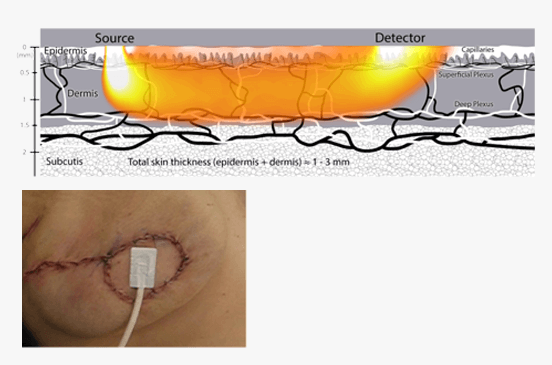
T.Ox sensor non-invasively placed on a breast reconstructive skin paddle.

I feel very comfortable with the dependability of T.Ox

Risal Djohan, MD

how can we help you?

Use this form to send your questions or feedback. We will get back to as soon as possible.

    I am Interested in

    FREE Clinical evaluationCustomer supportJob opportunitiesInvestor information

    * Required fields