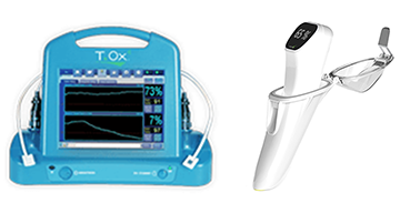
T.Ox
T.ox Remote
Intra.Ox
Intra.Ox Free Evaluation Now Available!
Standard of Care at Leading Hospitals
Numerous Clinical Studies
Alex Keller, MD
J. Brian Boyd, MD, FRCS, FRCSC, FACS
COUNTRY*USACanadaAfghanistanAlbaniaAlgeriaAmerican SamoaAndorraAngolaAnguillaAntarcticaAntigua and BarbudaArgentinaArmeniaArctic OceanArubaAshmore and Cartier IslandsAtlantic OceanAustraliaAustriaAzerbaijanBahamasBahrainBaker IslandBangladeshBarbadosBassas da IndiaBelarusBelgiumBelizeBeninBermudaBhutanBoliviaBosnia and HerzegovinaBotswanaBouvet IslandBrazilBritish Virgin IslandsBruneiBulgariaBurkina FasoBurundiCambodiaCameroonCanadaCape VerdeCayman IslandsCentral African RepublicChadChileChinaChristmas IslandClipperton IslandCocos IslandsColombiaComorosCook IslandsCoral Sea IslandsCosta RicaCote d'IvoireCroatiaCubaCyprusCzech RepublicDenmarkDemocratic Republic of the CongoDjiboutiDominicaDominican RepublicEast TimorEcuadorEgyptEl SalvadorEquatorial GuineaEritreaEstoniaEthiopiaEuropa IslandFalkland Islands (Islas Malvinas)Faroe IslandsFijiFinlandFranceFrench GuianaFrench PolynesiaFrench Southern and Antarctic LandsGabonGambiaGaza StripGeorgiaGermanyGhanaGibraltarGlorioso IslandsGreeceGreenlandGrenadaGuadeloupeGuamGuatemalaGuernseyGuineaGuinea-BissauGuyanaHaitiHeard Island and McDonald IslandsHondurasHong KongHowland IslandHungaryIcelandIndiaIndian OceanIndonesiaIranIraqIrelandIsle of ManIsraelItalyJamaicaJan MayenJapanJarvis IslandJerseyJohnston AtollJordanJuan de Nova IslandKazakhstanKenyaKingman ReefKiribatiKerguelen ArchipelagoKosovoKuwaitKyrgyzstanLaosLatviaLebanonLesothoLiberiaLibyaLiechtensteinLithuaniaLuxembourgMacauMacedoniaMadagascarMalawiMalaysiaMaldivesMaliMaltaMarshall IslandsMartiniqueMauritaniaMauritiusMayotteMexicoMicronesiaMidway IslandsMoldovaMonacoMongoliaMontenegroMontserratMoroccoMozambiqueMyanmarNamibiaNauruNavassa IslandNepalNetherlandsNetherlands AntillesNew CaledoniaNew ZealandNicaraguaNigerNigeriaNiueNorfolk IslandNorth KoreaNorth SeaNorthern Mariana IslandsNorwayOmanPacific OceanPakistanPalauPalmyra AtollPanamaPapua New GuineaParacel IslandsParaguayPeruPhilippinesPitcairn IslandsPolandPortugalPuerto RicoQatarReunionRepublic of the CongoRomaniaRussiaRwandaSaint HelenaSaint Kitts and NevisSaint LuciaSaint Pierre and MiquelonSaint Vincent and the GrenadinesSamoaSan MarinoSao Tome and PrincipeSaudi ArabiaSenegalSerbiaSeychellesSierra LeoneSingaporeSlovakiaSloveniaSolomon IslandsSomaliaSouth AfricaSouth Georgia and the South Sandwich IslandsSouth KoreaSpainSpratly IslandsSri LankaSudanSurinameSvalbardSwazilandSwedenSwitzerlandSyriaTaiwanTajikistanTanzaniaThailandTogoTokelauTongaTrinidad and TobagoTromelin IslandTunisiaTurkeyTurkmenistanTurks and Caicos IslandsTuvaluUgandaUkraineUnited Arab EmiratesUnited KingdomUSAUruguayUzbekistanVanuatuVenezuelaViet NamVirgin IslandsWake IslandWallis and FutunaWest BankWestern SaharaYemenYugoslaviaZambiaZimbabwe
How did you hear about us?GoogleWord of MouthSearch EngineSocial MediaAdvertismentFriendEventForum or BlogOther
I am Interested in
FREE Clinical evaluationCustomer supportJob opportunitiesInvestor information